


Which hospital is best for retina treatment in India?
There are several hospitals in India that offer excellent retina treatment, but one notable retina speciality hospital is Arohi Eye Hospital which is known for its advanced technology and experienced doctors. With state-of-the-art facilities and a patient-centric approach, Arohi Eye Hospital is a top choice for those seeking retina treatment in India.
How successful is retina surgery?
Retina surgery can be highly successful in treating conditions such as retinal detachment or macular holes. The success rate of the surgery can vary depending on factors such as the severity of the condition and the expertise of the surgeon. However, in most cases, the success rate is high, with a majority of patients experiencing significant improvement in their vision.
How many days after retina surgery can I see?
Full recovery of your best vision after retina surgery may take as long as six months or more. Vision will gradually improve as air or gas bubbles injected into the eye during surgery dissipate. Initially, your vision will be significantly blurred due to the bubbles. It is important to follow postoperative instructions and attend all follow-up appointments with your eye doctor.
Can a detached retina be repaired?
Yes, a detached retina can be repaired through surgery. The success rate of the surgery is high, with over 90% of detachments being repaired with one operation, though some people may require multiple surgeries. If the retina is not treated in time, it will also lead to vision loss.
What causes eye floaters?
The natural aging process of the eye typically causes floaters. Over time, the vitreous, a gel-like fluid in the eye, can clump together and create shadows on the retina, resulting in the appearance of floaters. Other factors, such as injury, inflammation, or certain medical conditions, can also cause floaters.
How long will my surgery last?
The duration of your surgery depends on the type of procedure and the complexity of your case. Your surgeon will provide you with an estimated duration before the surgery, and it typically ranges from 30 minutes to several hours. However, it’s important to note that the exact length of the surgery may vary depending on any unexpected complications that may arise during the procedure.
Will I need to use drops?
It depends on the type of surgery you are having and your individual needs. Drops may be necessary to prevent infection, manage pain, and promote healing. Your doctor will provide specific instructions on how to use the drops and any other post-operative care you may need. To ensure a successful recovery, it’s crucial to carefully follow these instructions.
When can I return to work?
The timeline for returning to work will depend on the nature and severity of your illness or injury. In general, it is recommended to take 2-4 weeks off to allow for proper recovery before returning to normal activities, including work.
How To Find the Right Retina Specialist in Your Area?
If you’re looking for a retina specialist in your area, a good starting point is to conduct a simple search using keywords such as “retina specialist near me” or “retina doctors near me.” This search can provide you with a list of the top specialists in your locality, making your search for the right doctor easier and more efficient.
What are retinal diseases, and why are they serious?
Retinal diseases affect the delicate tissue at the back of the eye, causing vision loss if untreated. Consulting a retina specialist in Mumbai ensures timely diagnosis and advanced retina treatment to preserve sight.
How are retinal tears or holes treated?
Retinal tears or holes are treated using laser photocoagulation or cryotherapy. A skilled retina surgeon in Mumbai performs these procedures to seal the retina and prevent detachment or further damage.
What treatments are available for diabetic retinopathy?
Diabetic retinopathy can be treated using laser therapy, injections, or vitrectomy treatment in Mumbai. An eye surgeon in Mumbai or the best retina specialist in Mumbai provides tailored retina treatment for vision restoration.
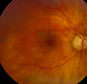
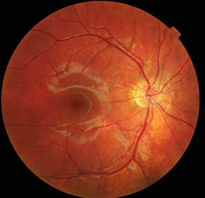
What diagnostic tools are used to evaluate retinal conditions?
Tools such as OCT, angiography, and fundus photography help assess retinal health. The best retina specialist in Mumbai uses these to detect abnormalities and plan precise retina treatment.
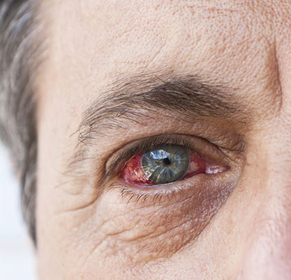
What are the early warning signs of retinal disease?
Symptoms like sudden floaters, flashes, or blurry vision may indicate retinal issues. Seek immediate care from a retina specialist in Mumbai for early detection and effective retina treatment in Mumbai.